
Fig. 5. TMEM24 functions as a lipid-transfer protein with a preference for PI.
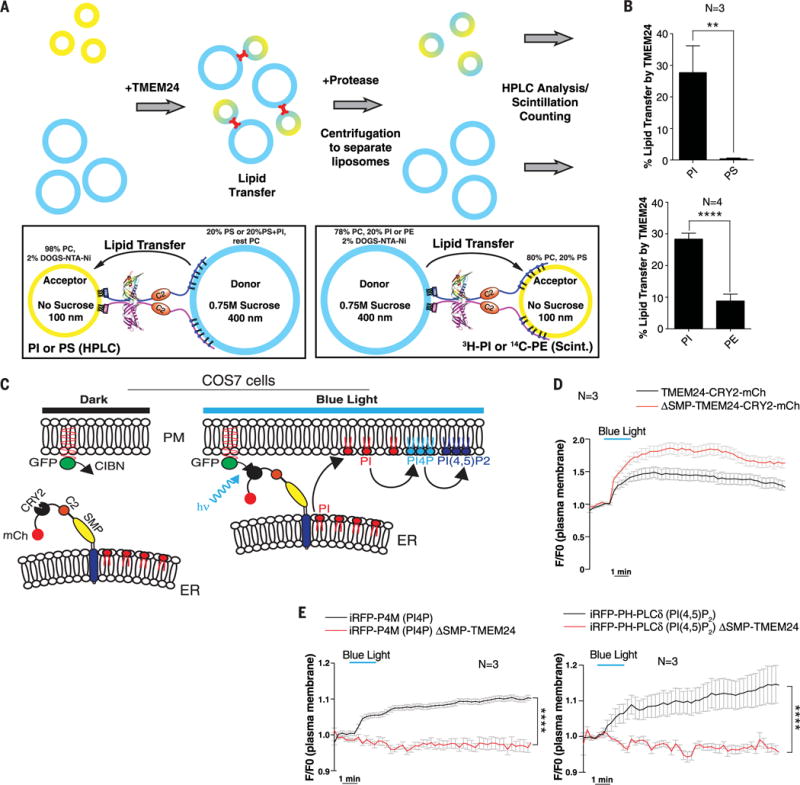
(A) In vitro lipid-transfer assays with TMEM24. (Top) Heavy donor (blue) and light acceptor (yellow) liposomes were mixed in the presence or absence of a recombinant N-terminally His12-tagged construct comprising the cytosolic portion of TMEM24 (residues 36 to 706). After lipid transfer (15 or 30 min),TMEM24 was degraded with protease and liposomes were separated by centrifugation. Donor and acceptor liposomes were separately analyzed for lipid content by HPLC or scintillation counting. (Bottom left) TMEM24 was tethered to donor liposomes containing PS via the basic C terminus and to acceptor liposomes containing 2 mole % (mol %) DOGS-NTA-Ni2+ via its N-terminal His12 tag. Transfer to acceptor liposomes of PS or PI, originally present at 20 mol % only in the donor liposomes, was measured by HPLC analysis after 30-min transfer. (Bottom right) TMEM24 was tethered to donor liposomes containing DOGS-NTA-Ni2+ via its N-terminal His12 tag and to acceptor liposomes containing PS via the basic C terminus. Transfer to acceptor liposomes of 14C-PE or 3H-PI, originally present only in the donor liposomes, was measured by scintillation counting after 15-min transfer. (B) Quantitation of the assays described in (A). TMEM24 preferentially transfers PI over PS (top panel) and PE (bottom panel). Mean ± SD indicated. (C) Schematic representation of light-induced TMEM24 PM recruitment strategy. TMEM24 was rapidly recruited to the PM by light-induced dimerization of CRY2 and CIBN modules. A version of TMEM24 with its C terminus replaced by a CRY2-mCherry module was expressed in COS-7 cells. The CIBN module is localized to the PM via a C-terminal CAAX box. (D) Dynamics of TMEM24-CRY2-mCherry (black) and ΔSMP-TMEM24-CRY2-mCherry (red) fluorescence before, during, and after blue-light stimulation (20 pulses, 200 ms each), as monitored by TIRF microscopy. (E) TIRF microscopy of PI(4)P marker iRFP-P4M (left) and the PI(4,5)P2 marker iRFP-PH-PLCδ (right) before, during, and after blue-light stimulation. Note the increase in both phosphoinositides due to transfer of PI to the PM by TMEM24 after blue-light stimulation (black). PM recruitment of ΔSMP-TMEM24-CRY2-mCherry does not affect PM levels of PI(4)P or PI(4,5)P2 (red). P values are P = 0.0049 and P < 0.0001 for (B) upper and lower panels, respectively, and P < 0.0001 for both (E) panels.