
Figure 2 (C).
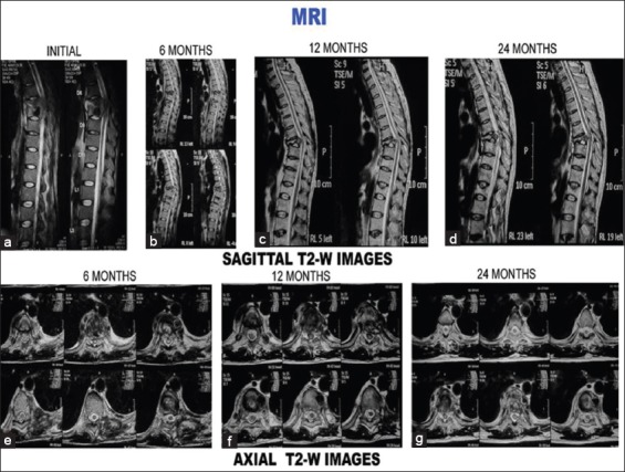
T2-weighted images (sagittal and axial) of the same patient. (a-d) Sagittal sections at presentation, 6 months, 12 months, and 24 months. (a) Initial sagittal sections show paradiscal lesion with endplate erosions, intraosseous caseation, marrow edema, epidural collection compressing spinal cord. All these features resolved subsequently. Disc is preserved although narrowed (b-d). (e) Axial sections at 6 months show septate paravertebral collection and intraosseous caseation. All these resolved subsequently (f-g)