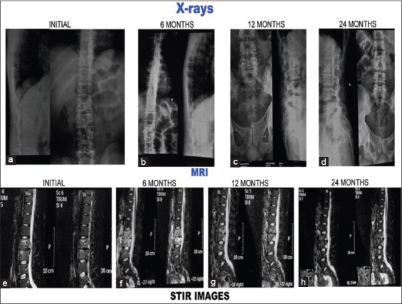
Figure 3 (A).

Plain X-rays and magnetic resonance imaging pictures of 20-year-old male with tubercular spondylitis L2–L3. Patient treated conservatively. Patient had associated polycystic kidney disease.(a-d): Plain X-rays at presentation, 6 months, 12 months, and 24 months. (a) Initial X-rays both anteroposterior and lateral show paradiscal lesion L2–L3 with endplate destruction, vertebral body destruction and narrow disc space. Endplate sclerosis occurred and lesion healed with narrow disc space (b-d). (e-h) short tau inversion recovery images of magnetic resonance imaging. (e) Initial short tau inversion recovery images show hyperintense signal intensity which subsequently decreased (f-g) to become isointense at 24 months (h)
