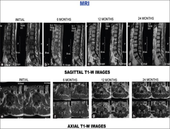
Figure 3 (B).

T1-weighted images (sagittal and axial) of the same patient. (a-d) Sagittal sections at presentation, 6 months, 12 months, and 24 months. (a) Initial sagittal sections show hypointense signal intensity with paradiscal lesion, discitis, endplate erosions, intraosseous caseation, and marrow edema. Six-month magnetic resonance imaging sections show increase in number of vertebrae involvement and increased edema (b). Healing occurred with increase in signal intensity on T1-weighted images (e-h) axial sections at presentation, 6 months, 12 months, and 24 months. (e) Initial axial sections show paravertebral collection, intraosseous caseation with mild epidural collection. All these resolved subsequently (f-h)
