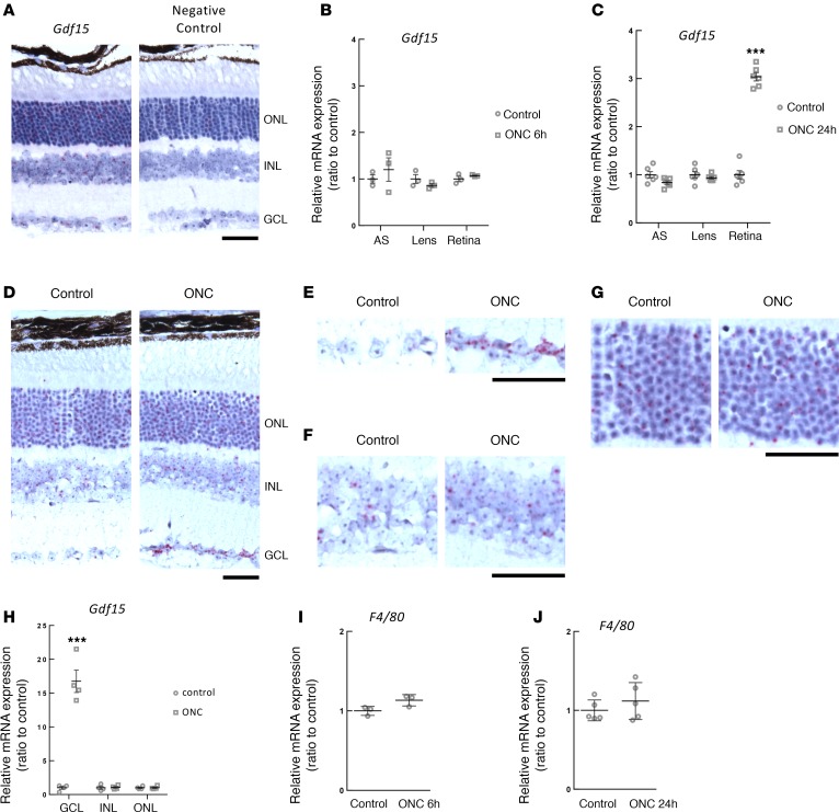
Figure 3. Ganglion cell layer (GCL) showed increased Gdf15 expression following axonal injury to the optic nerve.
(A) In situ hybridization of Gdf15 in the mid peripheral retina (n = 3 per group, representative pictures are shown). ONL, outer nuclear layer; INL, inner nuclear layer; GCL, ganglion cell layer. Scale bar: 50 μm. (B and C) Gene expression of Gdf15 in anterior segment (AS), lens, and retina (B) 6 hours (n = 3 per group) and (C) 24 hours after optic nerve crush (ONC) (n = 6 per group). (D) In situ hybridization of Gdf15 in the mid-peripheral retina 24 hours after ONC (n = 4 per group, representative pictures are shown). Scale bar: 50 μm. (E–G) High-magnification images from D. Scale bar: 50 μm. (E) In situ hybridization of Gdf15 24 hours after ONC in GCL. (F) In situ hybridization of Gdf15 24 hours after ONC in INL. (G) In situ hybridization of Gdf15 24 hours after ONC in ONL. (H) Gdf15 gene expression in GCL, INL, and ONL of the retina following the isolation by laser microdissection 24 hours after ONC (n = 4 per group). (I and J) F4/80 gene expression in the retina (I) 6 hours (n = 3 per group) and (J) 24 hours after ONC (n = 5 per group). Values are mean ± SD. ***P < 0.001 by 2-tailed unpaired t test.

