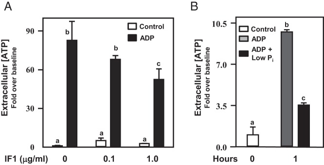
Figure 6. IF1 and low extracellular Pi inhibit extracellular ATP synthesis in αT3–1 cells.
Studies sought to regulate F0F1 ATP synthase activity at the cell surface in αT3–1 cells. In A, αT3–1 cells were treated with increasing doses of IF1 (0.1 and 1.0 μg/mL) and vehicle (open bars) or ADP (10μM; black bars) for a 1-hour period. Extracellular ATP levels were then assayed. IF1 blunted (P < .05) extracellular ATP synthesis in a dose-dependent manner. In B, similar studies were carried out examining the effects of reduced Pi in the absence or presence of ADP in the culture media. Media deficient in Pi could not support (P < .05) extracellular ATP synthesis in αT3–1 cells compared with media replete with Pi. Differing letters depicted over bars within a study designate statistical significance (P < .05).

