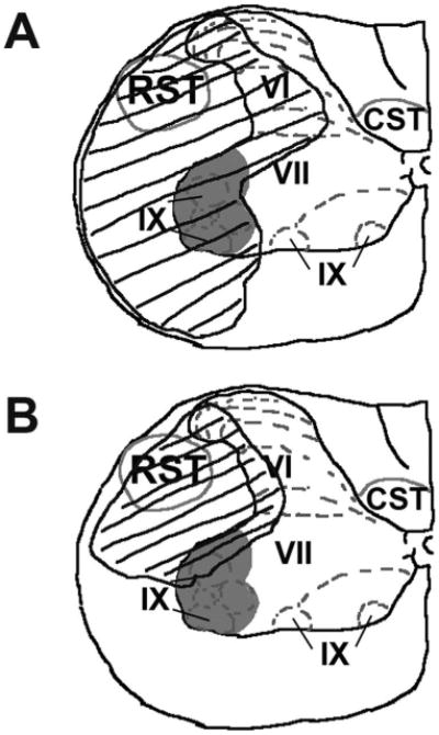
Figure 3.

Summary of damage in gray and white matter regions in epicenter of lesion in the ipsilateral spinal cord. The area of damaged tissue was traced in representative micrographs processed with cresyl violet and Luxol fast blue histochemistry using Neurolucida software from the SCI group (A) and the E2 5.0 group (B). The tracings were then fitted onto an anatomical atlas representation of the spinal cord at level C5 depicting motor neuron pools (gray shading) and white matter tracts (gray outline) associated with forelimb function (adapted from Kuchler et al., 2002). The damaged tissue area is depicted by the black outline and hashes. A reduction in damage in gray matter neuronal pools and white matter tracts associated with forelimb function was seen in the group that received post-SCI administration of 17β-estradiol. RST, rubrospinal tract; CST, corticospinal tract. Rexed's laminae VI, VII, and IX are indicated.
