Figure 4.

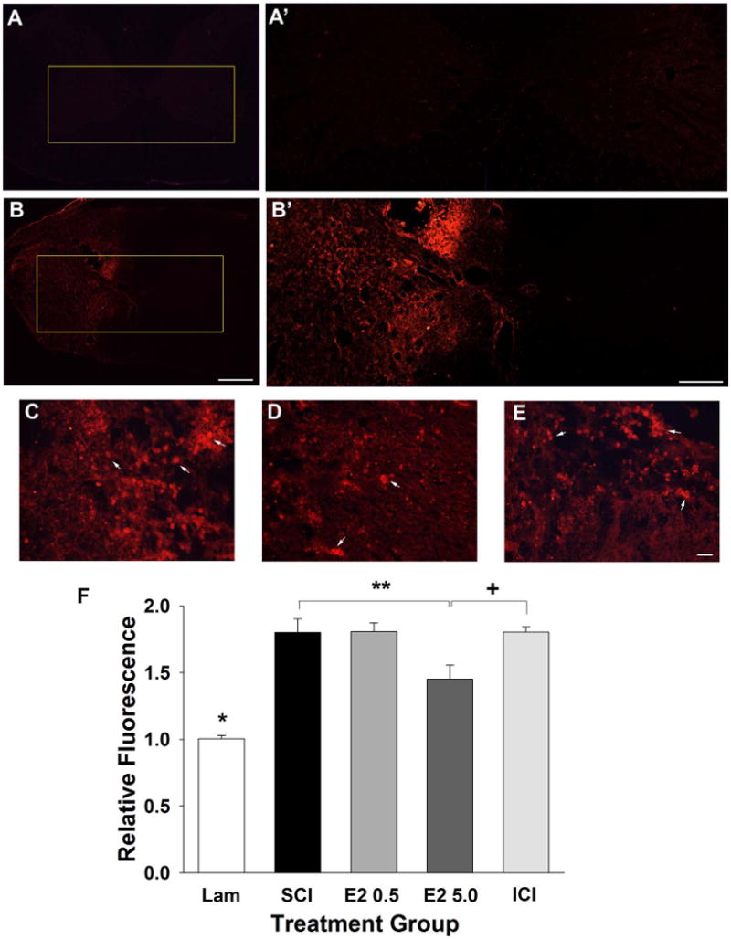
Effect of post-SCI administration of 17β-estradiol on microglial activation in the ipsilateral spinal cord. Microglial activation was evaluated by assessment of CD11B immunoreactivity and micrographs from representative transverse tissue sections at the epicenter of the lesion are shown from the uninjured group (A) and the SCI group (B). Higher magnification micrographs of the boxed regions are shown in A′,B′. Higher-magnification representative micrographs aligned by the central canal from the SCI (C), E2 5.0 (D), and ICI (E) groups demonstrate activated microglial cells (arrows) clustered throughout the ipsilateral spinal cord. Quantification of normalized relative fluorescence intensity of CD11b immunoreactivity is shown in F. The Lam group had a significantly lower relative fluorescence intensity than all SCI groups (indicated by *). A significant difference in fluorescence intensity was found between the SCI and E2 5.0 groups (indicated by **) and between the E2 5.0 and ICI groups (indicated by +). Scale bars = 400 μm in A,B; 200 μm in A′,B′; 20 μm in C–E. [Color figure can be viewed in the online issue, which is available at wileyonlinelibrary.com.]
