Figure 5.

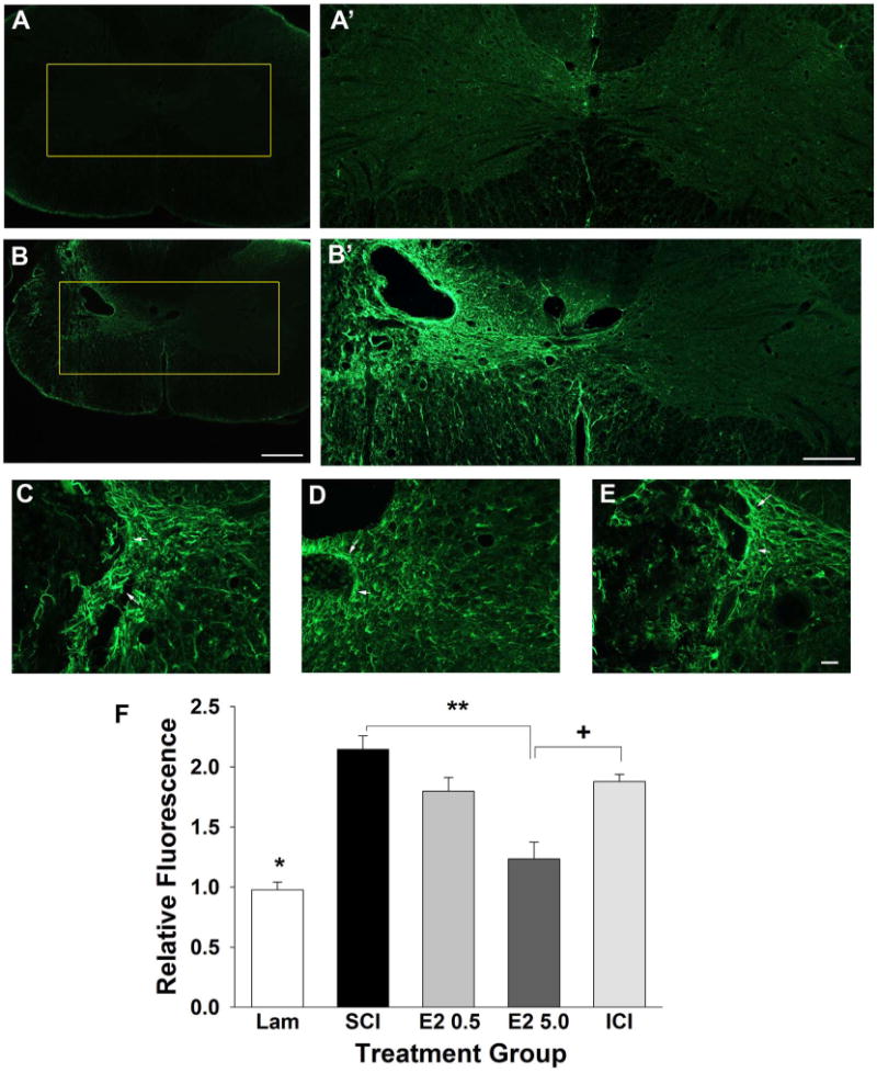
Effect of post-SCI administration of 17β-estradiol on reactive astrogliosis in the ipsilateral spinal cord. Reactive astrogliosis was quantified by GFAP immunoreactivity and micrographs from representative transverse tissue sections at the epicenter of the lesion are shown from the uninjured group (A) and the SCI group (B). Higher-magnification micrographs of the boxed regions are shown in A′ from the uninjured control group and B′ from the SCI group. Higher-magnification representative micrographs aligned by the central canal from the SCI (C), E2 5.0 (D), and ICI (E) groups demonstrate reactive astrogliosis (arrows). Quantification of normalized relative fluorescence intensity of GFAP immunoreactivity (F) demonstrated that the uninjured group had significantly lower intensity than all SCI groups (indicated by *). Also, we found that relative fluorescence intensity was significantly reduced in the E2 5.0 group as compared with the SCI group (indicated by **). The relative fluorescence intensity was significantly increased in the ICI group as compared with the E2 5.0 group (indicated by +). Scale bars = 400 μm in A,B; 200 μm in A′,B′; 20 μm in C–E. [Color figure can be viewed in the online issue, which is available at wileyonlinelibrary.com.]
