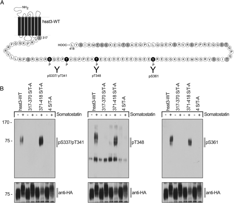
Figure 3. Generation of phosphosite-specific antibodies for the human sst3 receptor.
A, Schematic representation of the human sst3 receptor indicating all potential phosphate acceptor sites within the C-terminal tail in gray. Epitopes of the phosphosite-specific antibodies are depicted in black. B, HEK293 cells stably expressing wild-type hsst3, 317–370 S/T-A, 4S/T-A, or 371–418 S/T-A were either not exposed or exposed to 1μM SS-14 for 15 minutes at 37°C. The levels of phosphorylated sst3 receptor and phosphorylation-deficient mutants were then determined using the phosphosite-specific anti-pS337/pT341, anti-pT348, or anti-pS361 antibodies. Blots were subsequently stripped and reprobed with anti-HA antibody to confirm equal loading of the gels (anti-HA). Blots shown are representative of 3 independent experiments. The positions of molecular mass markers are indicated on the left (in kDa).

