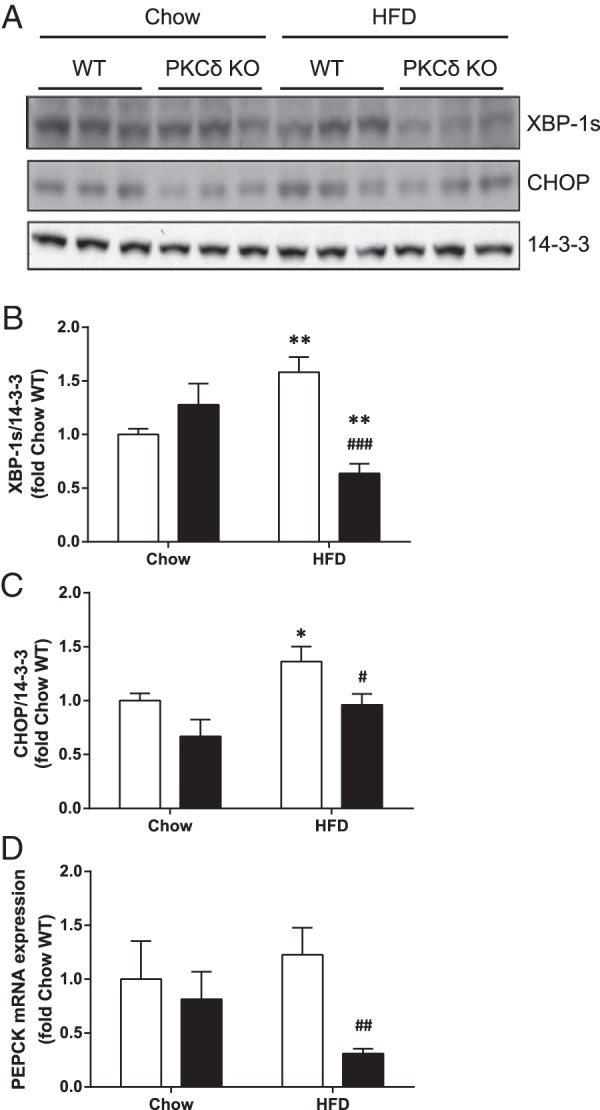
Figure 7. Effect of PKCδ deletion on ER stress markers and PEPCK gene expression in livers of fat-fed mice.

Liver lysates from chow or fat-fed WT (white bars) and PKCδ KO mice (black bars; n = 6 in each group) were subjected to immunoblotting to detect changes in XBP-1s and CHOP expression. Representative immunoblots (A) and mean ± SEM from densitometry (B and C) are shown. D, PEPCK mRNA expression in livers was analyzed by qRT-PCR. Student's t test; *, P < .05; **, P < .01 vs chow-fed mice; #, P < .05; ##, P < .01; ###, P < .001 vs HFD-fed WT mice.
