
Figure 5. Colocalization of UBE3C, ERα, and CCNB1 during mitosis.
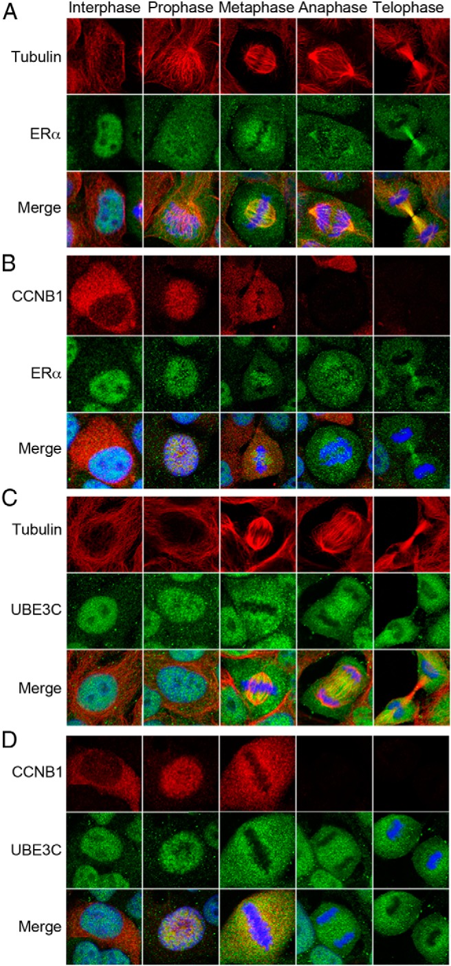
Exponentially growing MCF-7 cells were fixed, permeabilized, and coimmunostained with antibodies to either tubulin and ERα (A), CCNB1 and ERα (B), tubulin and UBE3C (C), or CCNB1 and UBE3C (D). The nucleus was stained with DAPI (blue). Merge indicates an overlay of the images of the 2 proteins and the nucleus. A representative cell in interphase and each phase of mitosis is shown.