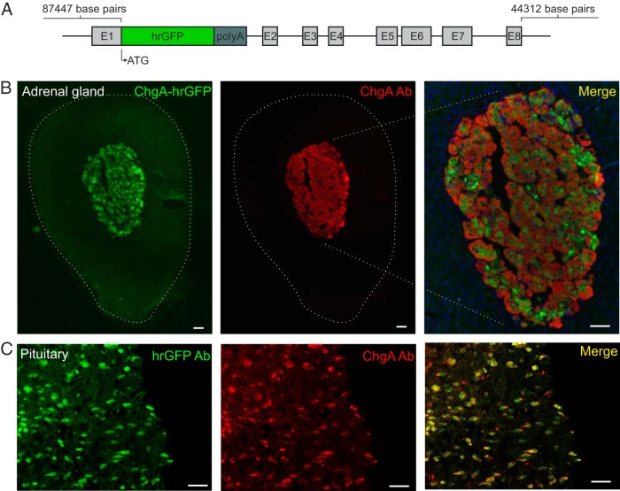
Figure 1. The ChgA-hrGFP construct and ChgA promoter-driven hrGFP expression in the adrenal medulla and pituitary gland.
A, The part of exon 1 after the start codon for ChgA was replaced with the coding sequence of hrGFP followed by a SV polyadenylation signal (polyA). Start codon (ATG) and exons for ChgA are indicated. B, Fluorescence microscopy showing ChgA promoter-driven hrGFP fluorescence (green) and ChgA immunoreactivity (red) in the adrenal gland (Ab from Abcam), and (C) the pituitary gland (Ab from Santa Cruz Biotechnology, Inc). The merged pictures also show DAPI nuclei staining (blue). Scale bars, 50 μm.

