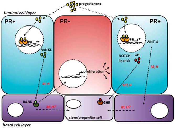
Figure 2. After P exposure, P enters a PR+ luminal cell (shown in blue) and binds to PR, which dimerises and transcribes target genes (eg, RANKL, WNT-4, NOTCH ligands, GH).
These signaling pathways then stimulate proliferation of neighboring PR− luminal cells (shown in red), potentially also involving cells in the basal cell layer (shown in purple). Not all of these pathways have been shown to be active in both the mouse (M) mammary gland as well as human (H) breast.

