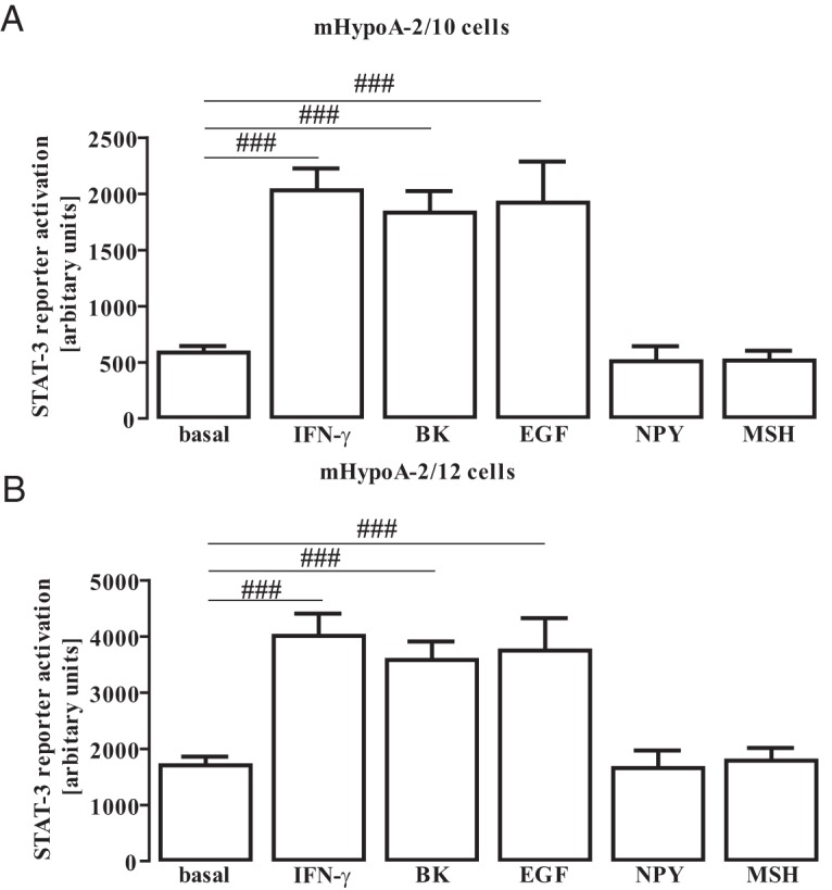
Figure 2. STAT-3 activation in hypothalamic cells.

A and B, mHypoA-2/10 (A) and mHypoA-2/12 (B) cells were transfected with a reporter gene construct containing 3 times the STAT-3 binding site (5′-TTCCCGTCAA-3′), serum starved for 16 hours, and then stimulated with IFN-γ (10 ng/mL), BK (1 μM), EGF (10 ng/mL), NPY (100 nM), or MSH (1 μM) for 6 hours. Data from 10 independent experiments performed in quadruplicate were compiled and are expressed as means ± SEM. Hash signs indicate a significant difference from basal.
