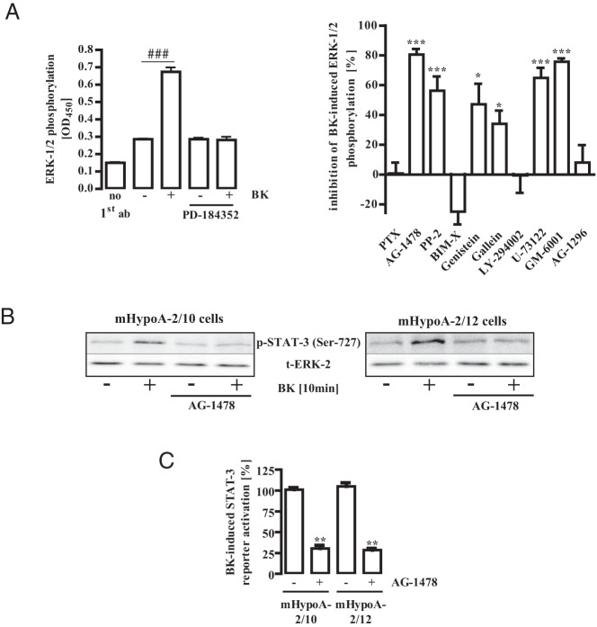
Figure 5. BK-induced ERK-1/2 and STAT-3 activation activity required EGFR activity.
A (left panel), mHypoA-2/12 cells were serum starved for 16 hours, preincubated for 30 minutes with PD-184352 (10 μM) or the carrier DMSO (0.1%), and then stimulated for 10 minutes with BK (1 μM). ERK-1/2 phosphorylation was detected by performance of a whole-cell ELISA with a p-ERK-1/2–specific antiserum. Data from 1 representative experiment performed in quadruplicate are given. Hash signs indicate significant differences from basal (−). A (right panel), mHypoA-2/12 cells were serum starved for 16 hours, preincubated for 16 hours with PTX (100 ng/mL), or for 30 minutes with BIM-X (1 μM), AG-1478 (100 nM), PP-2 (10 μM), genistein (50 μM), gallein (10 μM), Ly-294002 (20 μM), U-73122 (10 μM), GM-6001 (1 μM), AG-1296 (100 nM), or the carrier DMSO (0.1 and 0.2%), and then stimulated for 10 minutes with BK (1 μM). Inhibitor concentrations were chosen based on data from the literature, and inhibitors with no effects were tested in different assays. For example, PTX blocked the effects of NPY on cAMP and BIM-X blocked the effects of phorbol ester on ERK-1/2. ERK-1/2 phosphorylation was detected by performing a whole-cell ELISA with a p-ERK-1/2–specific antiserum. Data from 5 independent experiments performed in quadruplicate were compiled by calculating the inhibition as a percentage. Asterisks indicate a significant difference from 0%. B, mHypoA-2/10 or -2/12 cells were serum starved for 16 hours, preincubated for 30 minutes with AG-1478 (10 μM), and then stimulated for 10 minutes with BK (1 μM). Cell lysates were subjected to Western analysis using either a phospho-specific antiserum against Ser-727 of STAT-3 or against the total ERK-2 protein (t-ERK) to control for the total protein amount. One representative blot is shown. Data from 5 independent experiments are given in Table 1. C, mHypoA-2/10 or -2/12 cells were transfected with a STAT-3–dependent reporter gene construct, serum starved for 16 hours, preincubated for 30 minutes with AG-1478 (100 nM), and stimulated for 6 hours with 1 μM BK. The effects of AG-1478 on STAT-3 activation in 5 independent experiments performed in quadruplicate were compiled by defining ligand-induced reporter activation under basal conditions as 100%. Ligand-induced STAT-3 activation is expressed as the mean ± SEM, and asterisks indicate a significant difference from 100%.

