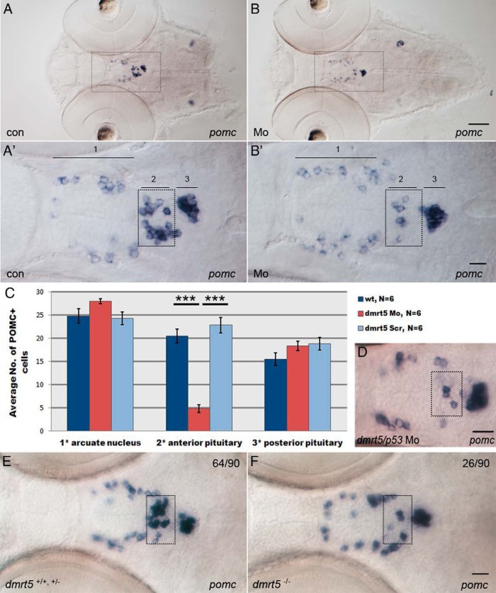
Figure 4. Knockdown of dmrt5 results in reduced corticotrope numbers.
A and B, Expression of pomc in control embryos (A) and dmrt5 morphants (B) at 72 hpf. A′ and B′ are higher-magnification views of areas boxed in panels A and B. pomc expression is shown in ventral diencephalon (arcuate nucleus, 1), corticotropes (anterior pituitary, 2 and boxed), and melanotropes (posterior pituitary, 3). C, Statistical analysis of pomc cell numbers showed that only corticotrope numbers (region 2) were significantly reduced (nMo = 4.8 ± 0.79, nwt = 20.5 ± 0.56, nctrMo = 22.8 ± 1; ***, P < .001). Error bars represent ± SEM and N is the number of analyzed embryos. D, Corticotrope defects could not be rescued by coinjection of p53 morpholino into dmrt5 morphants. E and F, pomc expression in wild-type and dmrt5ha2 mutant embryos at 72 hpf. Wild-type and heterozygous dmrt5 mutants had normal corticotrope numbers (E), whereas homozygous dmrt5 mutants phenocopied dmrt5 morphants and exhibited reduced corticotrope numbers (F). Numbers represent embryos with indicated phenotype in embryo clutch obtained from heterozygous parents. All images are ventral views; anterior is to the left. Corticotropes are boxed. Scale bars, 50 μm (A and B); 20 μm (A′–F).

