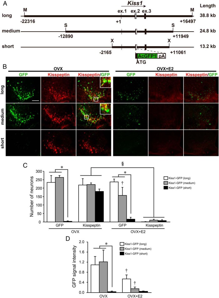
Figure 1. GFP reporter expression in the ARC of transgenic mice carrying three different lengths of GFP reporter constructs.
A, Schematic illustration of three constructs used for in vivo reporter assay. White boxes indicate the inserted AcGFP and polyA tail sequence. Positions and structures of these genes were obtained from the University of California, Santa Cruz, genome browser. Restriction enzyme recognition sites were indicated as follows: X, XhoI; S, SspI; M, MluI. B, Immunofluorescence analysis of kisspeptin and GFP expression in the ARC. Left and center panels show GFP and kisspeptin immunoreactivities in the ARC sections of representative transgenic mice, respectively, and right panels show computer-aided merged images of immunoreactive signals for kisspeptin (red) and GFP (green) in OVX or E2-implanted OVX (OVX+E2) mice. Insets show the sections at higher magnification. Scale bar, 100 μm. C, The number of GFP-expressing and kisspeptin-immunoreactive cells in the ARC of transgenic mice. D, The relative GFP signal intensity in the ARC of transgenic mice. Values are means ± SEM. *, P < .05 vs transgenic mice bearing long and medium-length constructs (two way ANOVA followed by Bonferroni test); †, P < .05 vs OVX mice bearing the same construct; §, P < .05 vs OVX mice as a main effect.

