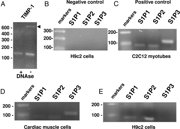
Figure 8. Expression of S1P receptor subtype in H9c2, primary cardiac muscle cells, and C2C12 myotubes.
A–E, Representative agarose gels of amplified cDNA fragments. Total RNA was incubated with DNase and successively reverse transcribed. cDNAs were then amplified by PCR using specific primers for TIMP-1 (A) and S1P1, S1P2, and S1P3 (B–E) as described in Materials and Methods. TIMP-1 amplification was performed by using untreated (−) or RNA treated with DNase (+) to eliminate DNA genomic contamination. B, Negative controls, consisting of no template (water) in each PCR. C, Positive controls consisting of cDNA obtained from murine C2C12 myotubes. D and E, cDNA amplification from reverse-transcribed RNA obtained from primary cardiac muscle (D) and H9c2 cells (E).

