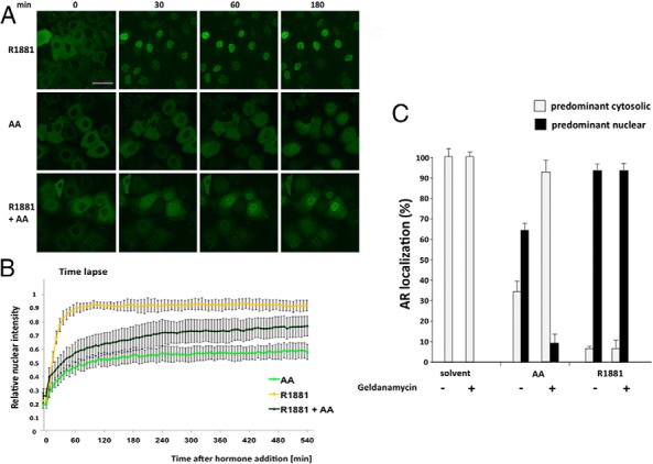
Figure 1. AA decelerates AR nuclear translocation in living cells: involvement of HSP90.

A, Confocal images of Hep3B cells stably expressing GFP-AR at different time points after treatment with R1881 (10−9 M), AA (10−4 M), or cotreatment. Scale, 50 μm. B, Live-cell time lapse imaging of Hep3B cells and quantification of GFP-AR nuclear translocation after addition of the indicated compounds. Calculation of the relative nuclear intensity (I) = [(Inucleus − Ibackground)/(Icytoplasm − Ibackground) + (Inucleus − Ibackground)]. The average of two experiments is shown, and the error bars indicate the variation of the mean of at least 25 cells per time point. C, The HSP90 inhibitor geldanamycin fosters the cytosolic localization of AR in the presence of AA. HeLa cells transfected with GFP-AR were treated for 2 hours with solvent, R1881 (10−10 M), or AA (3 × 10⁻⁵ M). Subsequently the HSP90 inhibitor geldanamycin (1 μg/mL) was added for 1 hour, and the intracellular localization of the AR was determined in 2 × 100 cells for each experiment. The deviation of the mean is shown.
