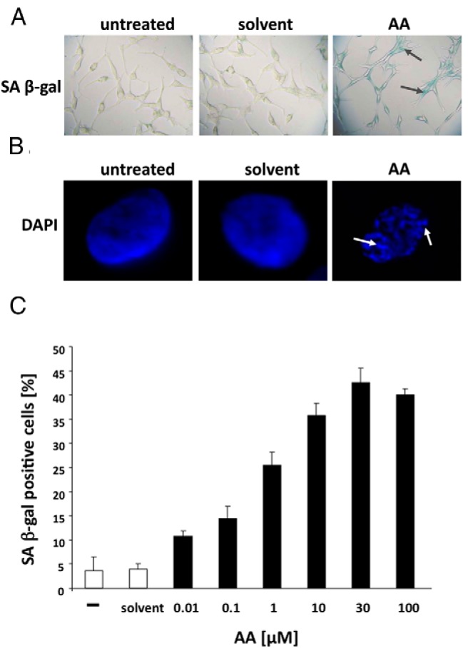
Figure 3. The natural AR antagonist AA induces cellular senescence in PCa cells.

Androgen-dependent growing LNCaP cells were treated with solvent control or the AR antagonist AA (3 × 10⁻⁵ M) for 72 hours. A, Phase-contrast microscope pictures of the staining for SA β-gal activity were taken at ×200 magnification. B, Fluorescence microscopy pictures at ×1000 magnification for the detection of SAHF via DAPI staining. SAHFs, as the marker of the induction of cellular senescence, are indicated by arrows. C, Quantification of SA β-gal-positive cells treated with the indicated concentrations. The 3 × 200 cells were counted and plotted as a percentage of positive cells. The error bars show the variation of the mean of triplets.
