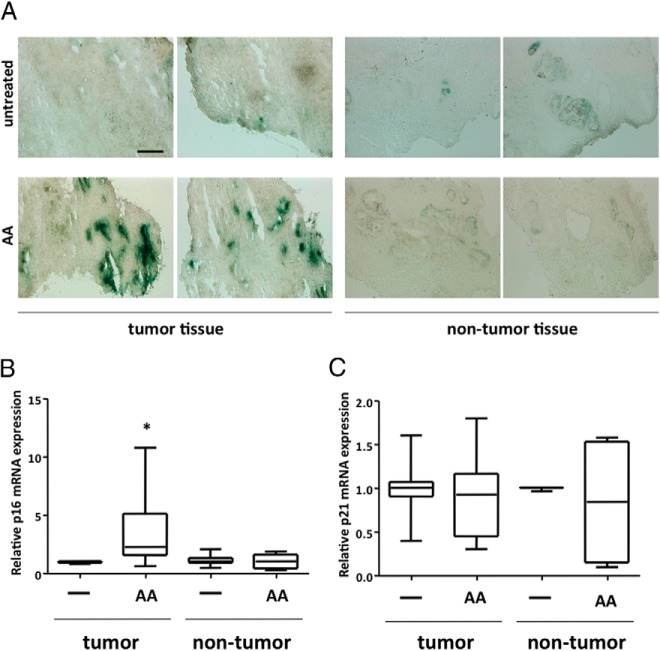
Figure 7. AA induces cellular senescence in human PCa tissues ex vivo.
Human PCa tissue (n = 6) and control/nontumor tissue (n = 3) specimens derived from prostatectomy of different patients (∼2 × 2 mm) were treated postoperatively for 48 hours with 10−4 M AA. A, Representative phase-contrast microscope pictures of SA β-gal staining of cryosections (10 μM thickness) of PCa tumor tissue or nontumor tissue. Scale, 300 μm. qRT-PCR was performed for p16 mRNA (B) and p21 mRNA (C). For internal normalization the mRNA expression of β-actin was determined. Box plot of relative p16 and p21 mRNA expression was used to compare the different treatment groups. Boxes show the lower and upper interquartile. Horizontal lines indicate the median and whiskers show a maximum of 1.5 interquartile range. Outliers were excluded.

