Figure 5.

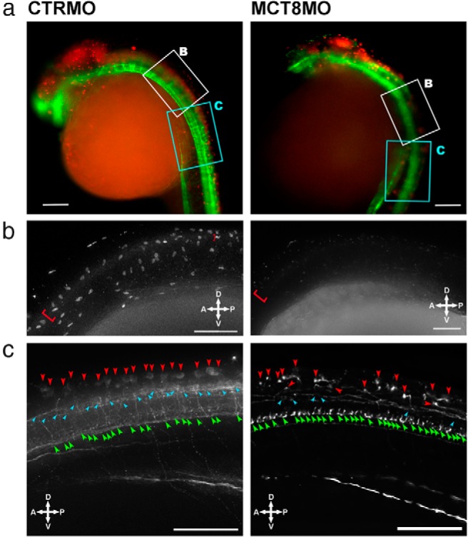
MCT8 knockdown affects brain and spinal cord development. A, Fluorescent images of 25-hpf wild-type zebrafish embryos microinjected with either CTRMO or MCT8MO. Red and green represent, respectively, Pax7 and acetylated β-tubulin immunostaining. The white and cyan boxed area denotes, respectively, the high-magnification images presented in B and C. Scale bars, 100 μm. B, Higher magnification of the most anterior spinal cord section of 25-hpf wild-type zebrafish embryos microinjected with either CTRMO or MCT8MO (white boxed area in A) after immunostaining for Pax7. The red brackets denote Pax7-positive spinal cord cells along the dorsal region of the developing spinal cord. In the MCT8 morphant, only the most anterior dorsal spinal cord has a few Pax7-positive cells. Scale bars, 50 μm. C, Higher magnification of sections 4 to 10 of the spinal cord in 25-hpf wild-type zebrafish embryos microinjected with either CTRMO or MCT8MO (white boxed area in A) after immunostaining for acetylated β-tubulin. Dorsal (red arrowheads), medial (cyan arrowheads), and ventral (green arrowheads) positioned spinal cord neurons are highlighted. Scale bars, 50 μm.
