Figure 6.
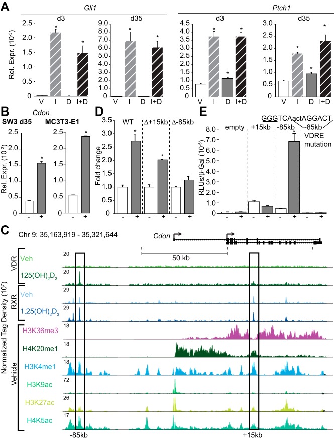
Regulation of the hedgehog pathway coreceptor encoding gene, Cdon, by 1,25(OH)2D3. A, Gene expression of cells treated with vehicle (V), 3 μg/mL IHH (I), 100 nM 1,25(OH)2D3 (D), or IHH + 1,25(OH)2D3 (I+D) were normalized to β-actin levels. Samples were analyzed in triplicate ± SEM (*, P < .05 vs vehicle). B, Cdon gene expression in IDG-SW3 and MC3T3–E1 cells treated with vehicle (−) or 100 nM 1,25(OH)2D3 (+) for 24 hours was normalized to β-actin levels and shown as fold change compared with d35 sample. Samples were analyzed in triplicate ± SEM (*, P < .05 vs vehicle). C, ChIP-seq tracks for Cdon, normalized to 107 tags, in the osteocyte (d35). D, BAC-Cdon-P2ALTN (WT, Δ+15kb, Δ-85kb) MC3T3–E1 stable cells treated with vehicle (−) or 100 nM 1,25(OH)2D3 (+) for 24 hours; relative light units (RLU) normalized to total protein and shown as fold change compared with vehicle ± SEM for a triplicate set of assays (*, P, .05 vs vehicle). E, pTK (empty), pTK-Cdon+15 kb, pTK-Cdon-85 kb region, or pTK-Cdon-85 kb VDRE mutated were transfected into MC3T3–E1 cells and treated with vehicle (−) or 100 nM 1,25(OH)2D3 (+). Each point represents relative light unit average normalized to β-gal ± SEM for a triplicate set of assays (*, P < .05 vs vehicle). Mutated sequence from GGG to AAA (underlined). d35, day 35; WT, wild type.

