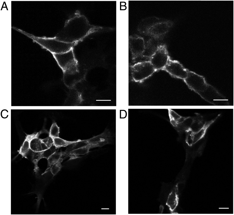
Figure 2.
Examination of the cell surface expression of the N321K-V2R. Immunofluorescence microscopy analysis of HEK293 cells transiently expressing WT (A and C) or N321K (B and D) HA-tagged V2R. The samples were stained with anti-HA-Alexa 488 mouse monoclonal antibodies under permeabilized (C and D) and nonpermeabilized (A and B) conditions. Scale bars corresponds to 10 μm.

