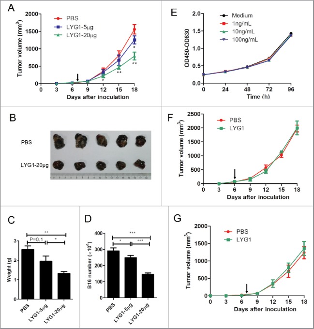
Figure 2.

Antitumor function of LYG1 is dependent on lymphocytes. (A–D) rhLYG1 inhibited B16 melanoma growth (n = 6–8 per group; one representative experiment was shown). B6 mice were inoculated s.c. with 2× 10⁵ B16 cells in the axilla, and rhLYG1 (5 μg or 20 μg) or PBS as a control was injected i.p. every day. Arrows represent the beginning of protein administration. (A) The tumor growth curve. (B) Excised tumors. (C) Tumor weights and (D) B16 cell numbers of excised tumors are shown. (E) CCK-8 assay of B16 cells with or without the addition of the indicated concentration of rhLYG1 in vitro. (F–G) B16 tumor progression following treatment with 20 μg rhLYG1 or PBS in SCID-beige mice (F) (n = 4 per group) or in Rag1−/− mice (G) (n = 5 per group). Arrows represent the beginning of protein administration. Results are representative of three independent experiments and expressed as the mean ± SEM, *p < 0.05 and **p < 0.01, LYG1–20 μg (or LYG1–5 μg) compared with PBS in Fig. 2A, LYG1–20 μg (or LYG1–5 μg) compared with PBS and LYG1–20 μg compared with LYG1–5 μg in Fig. 2C and D.
