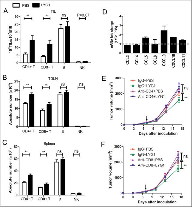
Figure 3.

T cells mediate the antitumor function of LYG1. (A–C) The absolute numbers of CD4+ T, CD8+ T, B, and NK cells in (A) TILs, (B) TDLNs, and (C) spleens in B16 tumor-bearing mice treated with 20 μg rhLYG1 or PBS as outlined in Fig. 2A, n = 6–8 per group. (D) Chemokines were examined by qPCR in excised tumors, n = 3 per group. Data were expressed as ratio of LYG1/PBS. More than one indicated upregulation upon LYG1 injection. (E and F) B16 tumor progression following treatment with 20 μg LYG1 or PBS and depletion of (E) CD4+ T cells or (F) CD8+ T cells. Arrows represent the beginning of protein administration, n = 6–8 per group. Results are representative of three independent experiments and expressed as the mean ± SEM, *p < 0.05 and **p < 0.01 compared with PBS.
