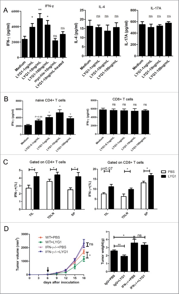
Figure 4.

LYG1 elicits CD4+ T cell-mediated tumor immunity by promoting IFNγ production. (A) IFNγ, IL-4 and IL-17A production by splenocytes treated with or without different concentrations of rhLYG1 and stimulated with coated anti-CD3 (1 μg/mL) and soluble anti-CD28 mAbs (0.5 μg/mL) for 48 h. Medium (LYG1 not added), Myc-6xhis peptide (10 ng/mL) and LYG1-heated (10 ng/mL) were used as negative controls. IFNγ, IL-4 and IL-17A concentrations in the culture supernatants of cells were measured by ELISA, and the results are shown on the vertical axis. (B) Naive CD4+ T cells or CD8+ T cells were isolated from LNs of WT B6 mice and stimulated with coated anti-CD3 (2 μg/mL) and soluble anti-CD28 mAbs (1 μg/mL). Various concentrations of rhLYG1 were added during the process. IFNγ concentrations in the culture supernatants of cells were measured by ELISA, and the results are shown on the vertical axis. (C) Single-cell suspensions isolated from TIL, TDLN, and spleen (SP) of tumor-bearing mice treated with 20 μg LYG1 or PBS were stimulated with PMA and ionomycin for 5 h and then tested for the expression of IFNγ by flow cytometry, n = 6–8 per group. The percentages of IFNγ+ cells gated on CD4+ T cells (left) and CD8+ T cells (right) are shown on the vertical axis. (D) B16 tumor progression and weights in WT and Ifng−/− mice treated with 20 μg LYG1 or PBS, n = 4–5 per group. Arrows represent the beginning of protein administration. This panel shows a representative statistical result of three independent experiments. Data are expressed as the mean ± SEM, *p < 0.05 and **p < 0.01 compared with medium or PBS.
