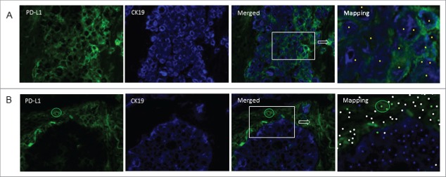
Figure 3.
PD-L1 expression on tumor and or stromal/immune cells. Frozen tissue sections derived from patients with lung adenocarcinoma were double-stained by immunofluorescence (CK19 (blue), PD-L1 (green). Tissue segmentation was performed on the basis of CK19 staining to determine tumor nest and stromal areas. A phenotyping step based on “training” of the software to recognize positive and negative cells was then performed generating an analysis algorithm. Mapping determined the phenotype of the cells (CK19+PD-L1−: blue dot; CK19+PD-L1+: yellow dot; CK19−PD-L1+: green dot; other cells: white dot). Positive (A) and negative (B) staining of tumor cells for PD-L1 is shown. The circle in B identifies PD-L1-positive cells in the stroma (original magnification ×200).

