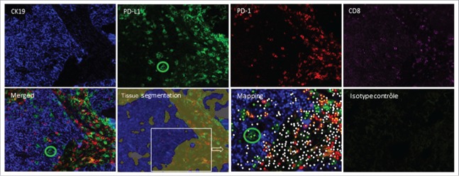
Figure 5.
Difficulties in the interpretation of PD-L1 staining in the tumor nest. Frozen tissue sections derived from patients with lung adenocarcinoma were quadruplex stained by immunofluorescence (CK19 (blue), PD-L1 (green), PD-1(red) and CD8 (pink)) on the same slide. Tissue segmentation was performed on the basis of CK19 staining to determine tumor and stromal areas. Mapping determined the phenotype of the cells with the corresponding code: CK19+PD-L1−: blue dot; CK19+PD-L1+: yellow dot; CK19−PD-L1+: green dot, PD-1+CD8+: orange dot; PD-1−CD8+: pink dot, PD-1+CD8−: red dot, other cells: white dot). The green circle identifies PD-L1+CK19− cells infiltrating the tumor nest (original magnification ×200).

