
Figure 5.
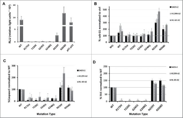
Activity in tumor cells of wild-type and mutant p53. (A) p53 activity levels in cancer cells were measured using a cis-reporter system. H1299-A2 cells were transfected with p53-Luc plasmid and electroporated 4 h later with the different p53 mutants RNAs. 48 h later, luciferase activity was measured using a luminometer and is presented herein as relative light units (RLU). pFC-p53 used as positive control plasmid for p53 expression. (B–D) Influence of various p53 proteins on the levels of subG1 population, Caspase 3 and FAS expression. Cells were electroporated with mRNAs encoding different p53 mutants. 48 h later, cell cycle (B), expression levels of the pro-apoptotic protein caspase-3 (C) and FAS receptor levels (D) were analyzed by flow cytometry. Data shown as a percentage of SubG1/Caspase-3/FAS levels, normalized to control (as mean ± SEM; n = 4).