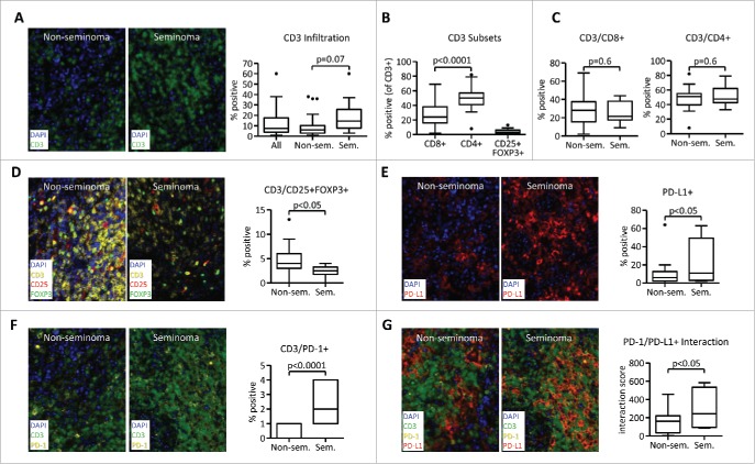
Figure 1.
Seminoma histology is associated with decreased Treg cell infiltration and higher PD-1/PD-L1 interaction. Samples from a cohort of 34 testicular germ cell tumor patients were analyzed using Quantitative Multiplexed Fluorescence Immunohistochemistry with Automated Quantitative Analysis (AQUA). Samples were stained with DAPI and antibodies against CD3 (A) or CD3 in combination with CD4, CD8, CD25, and FOXP3 (B–D). (E) Expression of PD-L1 in the tumor microenvironment. (F) Percentage of PD-1+ cells as a ratio from CD3+ DAPI+ cells. (G) PD-1/PD-L1 interaction scores. For data quantification, percentage of all DAPI+ nuclear cells (for CD3 and PDL1) or percentage from parent CD3 population (CD4+, CD8+, and CD25+FOXP3+) was used. PD-1/PD-L1 interaction is a numerical representation of the proportion of PD-1 positive cells within a pre-defined distance to PD-L1. The value is normalized by the total number of DAPI+ cells. All images were obtained using 20× zoom and were scaled digitally. In all images, 1 pixel = 0.5 µm.

