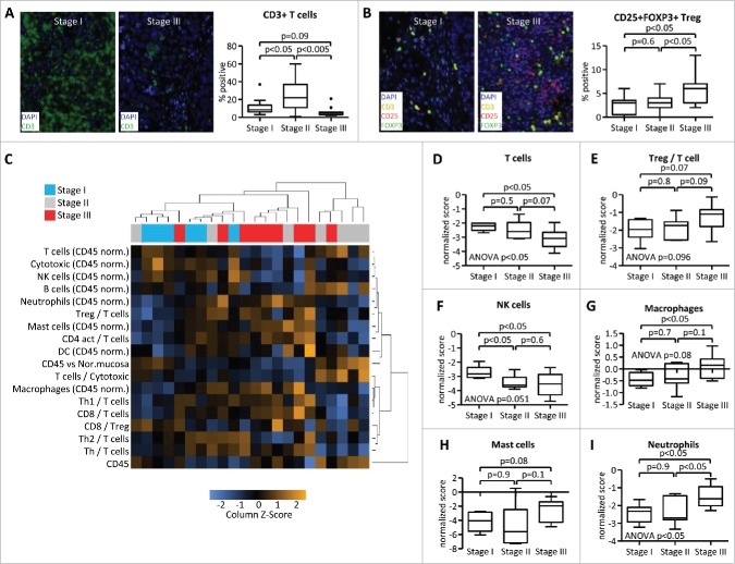
Figure 4.
Advanced stage is associated with changes in immune cell infiltration. Samples from testicular germ cell tumors were analyzed by Quantitative Multiplexed Immunohistochemistry (A, B) and by gene-expression profiling (C–I). Stage I (localized disease), stage II (lymph node metastases), and stage III (disseminated metastases) tumors were analyzed for CD3 expression (A) and CD3/CD25/FOXP3 co-expression (B). Numbers represent percentages of all DAPI+ nuclear cells (A) or of all CD3+ cells (B). (C) Gene-expression data were subjected to unsupervised clustering of both patient samples and scores of selected immune signatures. (D–I) Immune gene signature scores of stage I/II/III tumors were calculated by nSolver software.

