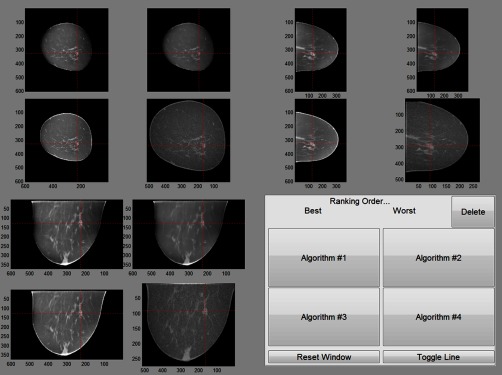
Fig. 4.
The layout of the viewer used for the second reader study. Each cross-sectional view of four selected reconstructions was grouped. Target lesion was highlighted. For this reader study, radiologists reviewed the center slice of each view of four selected reconstructions and ranked them in terms of which reconstruction provided the best diagnostic information (or simply their preference of one reconstruction algorithm over others).

