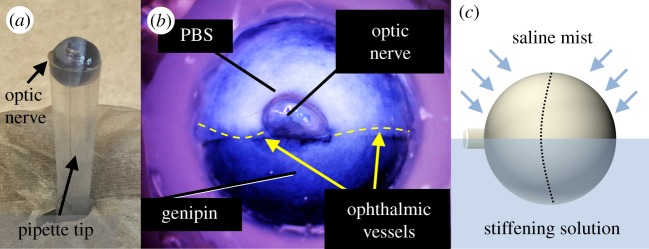
Figure 1.
Eyes were partially immersed in cross-linking agents, exposing approximately half the eye to a stiffening agent overnight by mounting it in a trimmed pipette tip (a). Genipin, which is also used as a blue dye, provides a visual indicator of its location (b). This is closely localized to the treated region and demonstrates little evidence of wicking. Regions appearing blueish near the top of panel (b) are actually thin regions of translucent sclera where choroid is visible, not regions exposed to genipin. Eyes were then incubated overnight while misting the tissue-draped control half with PBS to keep it moist (c). Dashed line indicates the limbus.

