Figure 6.

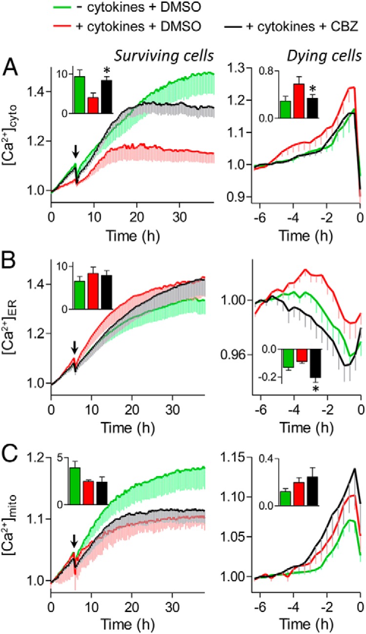
Carbamazepine (CBZ) modulates cytosolic, ER, and mitochondrial Ca2+ signaling. Dispersed mouse islet cells were transfected with D3cpv (A), D1ER (B), or 4mtD3cpv (C) genetically encoded Ca2+ biosensors to image cytosolic, ER, or mitochondrial Ca2+, respectively. Cells were incubated in 20 mM glucose, serum-free RPMI 1640 medium before the treatments with cytokines (red), cytokines and 100 μM carbamazepine (black), or no cytokines (green) at the indicated time (arrow). Left, Ca2+ levels of cells that remained alive during the 38 hours of imaging. Inset, area under the curve from the time of treatment to the end of the time course (n = 6–28 cells; ✻, P < .05 compared with cytokine treated). Right, Ca2+ levels of cells that died during the 38 hours of imaging. The FRET/cyan fluorescent protein ratios were normalized to the average reading from 5 to 6 hours before PI incorporation (0 hours). Inset, area under the curve from the time of Ca2+ influx or depletion to the time of PI incorporation (n = 11–26 cells; *, P < .05 compared with cytokine treated).
