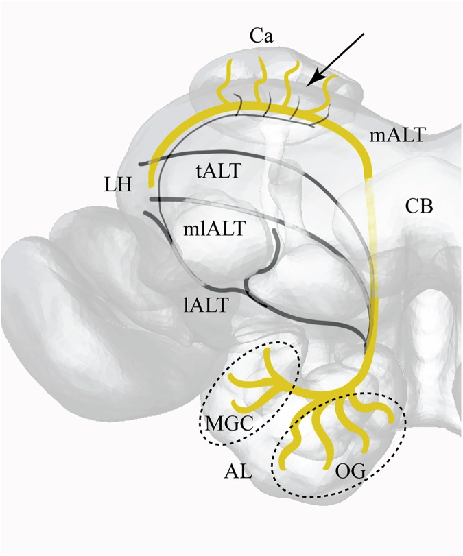
Fig 1. Projection view of a 3D-model of the right hemisphere with antennal lobe tracts and major brain areas indicated.
Arrow indicates insertion point for fluorescent dye into the calyx. AL (Antennal Lobe), Ca (Calyx), CB (Central Body), LH (Lateral Horn), mALT (medial AL tract), tALT (transverse AL tract), mlALT (mediolateral AL tract), lALT (lateral AL tract), MGC (Macro Glomerular Complex), OG (Ordinary Glomeruli).

