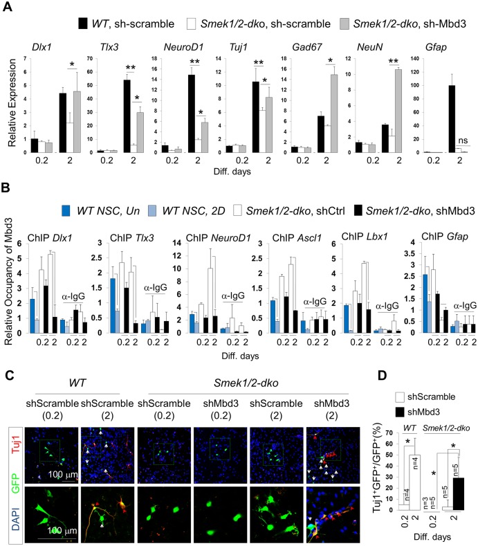
Fig 7. Effect of methyl-CpG–binding domain protein 3 (Mbd3) knockdown on neuronal gene expression and promoter occupancy over the course of differentiation of Suppressor of Mek null double knockout (Smek dKO) neural progenitor cells (NPCs).
(A) Wild-type (WT) or Smek1/2 dKO NPCs were electroporated with either control pLKO3G-shScramble or pLKO3G-shMbd3 lentiviral vector and grown for 2 d in N2 medium with basic fibroblast growth factor (bFGF). qPCR analysis was performed to detect indicated mRNAs (n = 3 or 6). (B) Mbd3 knockdown in Smek1/2 dKO NPCs decreases Mbd3 occupancy of Dlx1, Tlx3, NeuroD1, Ascl1, and Lbx1 promoters but not that of Gfap, as determined by chromatin immunoprecipitation-quantitative PCR (ChIP-qPCR) (n = 3). (C) Immunostaining to detect Tuj1 (red) and enhanced green fluorescent protein (EGFP) (green) expression. Nuclear staining is shown by 4',6-diamidino-2-phenylindole (DAPI) (blue). Scale bar, 100 μm. Red arrows indicate Tuj1/EGFP double-positive cells, and white arrows indicate EGFP-positive cells. (D) Quantification of panel C. All data are presented as average ± SD. t test analysis was performed to calculate significance (*p < 0.05, **p < 0.005; not significant (ns), p > 0.05). All individual quantification data underlying panels A, B, and D can be found in S2 Data.

