FIGURE 3:
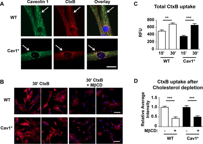
Endocytosis of CtxB. (A) Human fibroblasts were incubated for 30 min with 7.5 μg/ml Alexa 594–labeled CtxB at 37°C in the presence of 0.1 mg/ml BSA, fixed, and stained for Cav1 (green). Similar colocalization and uptake were evident in both control and Cav1* fibroblasts. Scale bar, 10 μm. (B–D) Uptake of CtxB over time is similar in WT and Cav1* human fibroblasts, and cholesterol depletion by 5 mM MβCD similarly reduced CtxB uptake in WT and Cav1* human fibroblasts. For the time course, images from five independent coverslips were obtained for each group, and fluorescence intensities were analyzed by tracing the cell contour (>100 cells/group). For CtxB uptake analysis, images from eight independent coverslips were analyzed for each group (>200 cells/group). Scale bar, 100 μm.

