FIGURE 4:
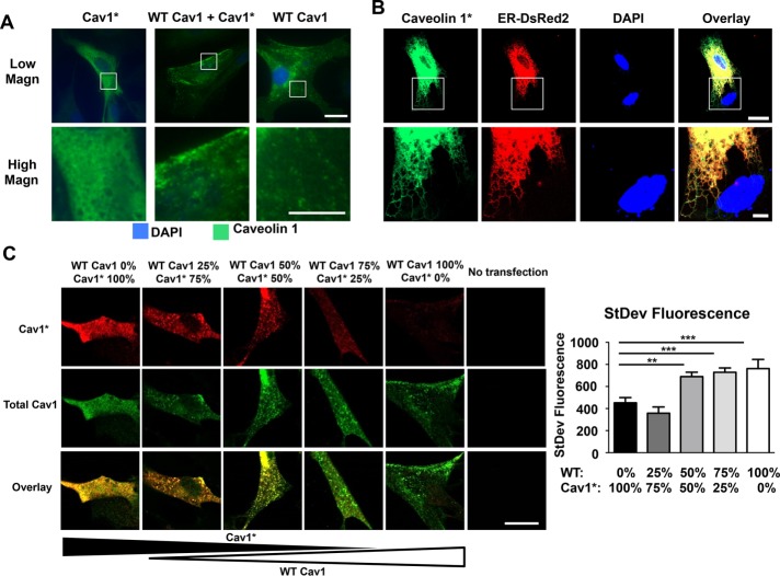
Dependence of caveolae formation on varying WT Cav1 and mutated Cav1* ratios. (A) Fibroblasts derived from Cav1−/− mice were transfected with either human WT Cav1 or human mutated Cav1*, followed by immunofluorescence analysis. Punctate staining was seen in fibroblasts expressing WT Cav1, whereas the pattern was diffuse after expression of Cav1*. On carrying out double transfection with both plasmids, fibroblasts showed relatively normal punctate staining pattern. Scale bar, 20 μm (top), 10 μm (bottom). (B) High-magnification view of Cav1−/− fibroblasts cotransfected with Cav1* and p-DsRed2-ER, showing localization of Cav1* in ER. Scale bar, 20 μm (top), 5 μm (bottom). (C) Cav1−/− fibroblasts were transfected with different ratios of WT Cav1 and Myc-tagged Cav1* (total amount of plasmid DNA was kept constant at 1 μg of plasmid DNA/150,000 cells), and the cells were stained with an antibody against Myc (red) and total Cav1 (green). Puncta formation was assessed by measuring the SD of the green fluorescent signal. Diffuse staining was seen in cells expressing only Cav1*, whereas higher expression of WT Cav1 led to a more heterogeneous and punctate staining. We also observed mutant Cav1* in puncta, as shown by yellow staining in cells transfected with a 1:1 ratio of Cav1 and mutant Cav1*. A higher-magnification view is shown in Supplemental Figure S4C. Scale bar, 20 μm.

