FIGURE 7:
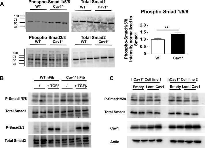
Increased Smad1/5/8 signaling in Cav1* fibroblasts. (A) Immunoblot analysis in control and Cav1* fibroblasts showing Smad1/5/8 hyperphosphorylation and normal Smad2/3 phosphorylation in Cav1* fibroblasts. Proteins were isolated from cells grown in DMEM plus 10%FBS. (B) Stimulation with 10 ng/ml TGFβ for 1 h similarly increased Smad2/3 phosphorylation in both cell types, confirming that signaling downstream of TGFβ is normal both at baseline and after stimulation. (C) To rescue WT Cav1 expression in human Cav1* fibroblasts, we transduced two different human Cav1* fibroblast cell lines at MOI 5, leading to doubling of Cav1 levels. We did not observe a change in phosphorylation of Smad1/5/8 in response to WT Cav1 up-regulation, arguing against haploinsufficiency as an explanation for increased phosphorylation of Cav1* patient fibroblasts.

