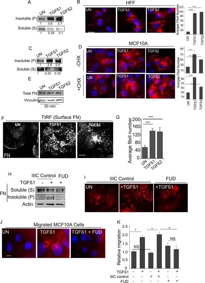
FIGURE 1:
TGF-β1 and TGF-β2 rapidly increase fibrillogenesis. (A, B) HFFs were treated with TGF-β1 or TGF-β2 for 30 min and (A) DOC extraction used to fractionate DOC-soluble (S) and -insoluble (pellet (P)) pool and immunoblotted for FN, or (B) cells were immunostained using anti-FN. Right, average fibril number (Materials and Methods) in untreated (UN), TGF-β1–, or TGF-β2–treated cells. Fibril lengths were tracked using the NeuronJ plug-in on ImageJ. (Materials and Methods). (C) MCF10A cells treated with TGF-β1 or TGF-β2 for 30 min and processed for DOC extraction and immunoblotted for (S) and (P) FN as in A. (D) MCF10A cells untreated or treated with 10 ng/ml TGF-β1 or TGF-β2 for 30 min (with or without preincubation with 20 µg/ml CHX for 2 h) and immunostained for endogenous FN. Right, average fibril number in untreated (UN), TGF-β1–, or TGF-β2–treated cells. Fibril lengths were tracked using the NeuronJ plug-in on ImageJ. Scale bar, 5 μm. (E) Total MCF10A cell lysates at the indicated treatment times were lysed in SDS buffer to solubilize total FN pools ((S) and (P) combined) and immunoblotted against FN. Vinculin was used as the loading control. (F) Cells treated as in A and imaged using TIRF microscopy (penetration depth, 110 nm). Scale bar, 5 μm. (G) Average fibril number analyzed from TIRF images in untreated (UN), TGF-β1–, or TGF-β2–treated cells. Fibril lengths from F were tracked using the NeuronJ plug-in on ImageJ. (H, I) MCF10A cells were treated with 10 ng/ml TGF-β1 for 30 min (with or without 500 nM FUD peptide or the matched control IIIC peptide) and processed either (H) for DOC extraction and immunoblotting for (S) and (P) FN (actin was the loading control from the soluble pool) or (I) immunostained for FN. (J) MCF10A cells allowed to migrate for 6 h in the presence of Rh-FN as indicated in the figure. Migrated cells are captured by fixing the cells on the Transwell filter. Images of Rh-FN are representative of at least four different fields on the filter from two independent biological trials. Scale bar, 5 µm. (K) Transwell migration through FN-coated Transwells of MCF10A for 12 h either untreated or in the presence of TGF-β1 alone or with either control IIIC peptide or FUD peptide as in J and as indicated. Migrated cells were counted and plotted relative to untreated filters. Asterisks indicate significant differences as indicated (*p < 0.05, **p < 0.01, ***p < 0.001). Quantitation of blots is representative of a minimum of three independent trials.

