FIGURE 2:

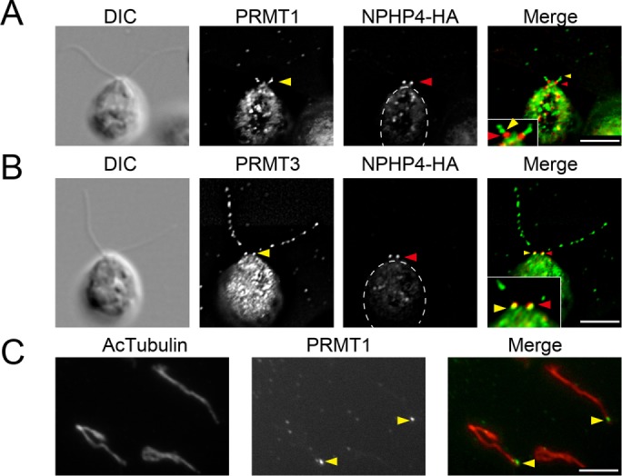
PRMT 1 and 3 localization at the base of flagella. (A, B) Immunofluorescence microscopy using cells expressing HA-tagged NPHP4 detected with anti-HA antibody (red). The cells are also labeled (green) with anti-PRMT 1 (A) and anti-PRMT 3 (B). Yellow arrowheads indicate the basal localization of PRMTs. Red arrowheads indicate the location of NPHP-HA. The basal localization of PRMT 1 is more distal than that of NPHP4-HA, a marker of the transition zone. By comparison, a portion of the PRMT 3 signal colocalizes with the NPHP4-HA signal. Insets, expanded images of the flagellar base. (C) Immunofluorescence microscopy of isolated flagella. Flagella are stained with anti–acetylated tubulin (Ac-tubulin; red) and anti-PRMT 1 (green). Localization of PRMT 1 is still detected at the base of the detached flagella, confirming the localization of PRMT 1 at a position distal to the transition zone. Cell bodies are outlined with dashed lines. Scale bars, 5 µm.
