
Figure 4.
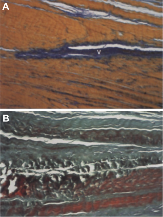
Comparison of histologic appearance with normal and degenerative coracoacromial ligament. (A) Normal appearance with parallel collagen bundles and elastic fibers. (B) Degenerative changes with loss of fiber orientation, decreased cell number, and overall disarray. V, blood vessel. (Reprinted with permission from Panni et al.46)