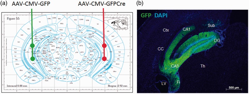
Figure 1.
Experimental design of brain-derived neurotrophic factor depletion.
(a) WT (BDNFf/f: 5XFAD−/−) and 5XFAD (BDNFf/f: 5xFAD+/−) mice were injected in the CA1 and CA3 regions (Bregma coordinates −2.9 mm A/P, ± 2.8 mm M/L, −2.5 and −4.0 mm D/V) of the HC with two 1 -µL injections of 1 × 10⁹ GC/µL AAV9-CMV-GFP virus into the left HC, and two 1 -µL injections of 1 × 10⁹ GC/µL AAV9-CMV-GFP-Cre virus into the right HC. (b) Representative section showing GFP fluorescence localized to the HC at 4 weeks after injection into a BDNFf/f: 5XFAD−/− mouse with AAV-CMV-GFP.
Ctx = cortex; LV = lateral ventricle; Th = thalamus; Sub = subiculum; Fi = fimbria of the hippocampus; CC = corpus callosum; DG = dentate gyrus.

