Figure 5.
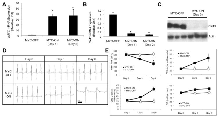
c-MYC gain-of-function in cardiomyocytes results in cardiac conduction defects that phenocopy BRG1/BRM loss-of-function. (A) RT-qPCR analysis of c-MYC mRNA levels in the heart of an inducible transgenic mouse line prior to induction (MYC-OFF) and 24–48 hours after DOX-mediated induction to overexpress c-Myc (MYC-ON, Day 1 and Day 2). Data are normalized to Gapdh and presented as means ± SEM based on 5 independent experiments with significant differences indicated (*p<0.05). (B) RT-qPCR analysis of Cx43 mRNA levels in heart of the same transgenic mouse line. Data are normalized to Gapdh and presented as means ± SEM based on 5 independent experiments with significant differences indicated (*p<0.05). (C) Representative western blot of CX43 protein levels in heart of same transgenic mouse line prior to induction (MYC-OFF) and after DOX-mediated induction (MYC-ON, Day 3). Actin serves as a loading control. 3 independent samples for MYC-OFF and MYC-ON are shown. (D) ECG sample trace readings from 3 MYC-OFF controls and 4 MYC-ON mice showing Wenckebach second-degree heart block by 3 days of DOX-induced c-MYC induction and a complete heart block by day 6. (E) Four panels showing ECG-based measurements from 3 MYC-OFF controls and 4 MYC-ON mice at three time points relative to DOX-mediated induction. The plots show significant differences (*p<0.05) in heart rate, PR interval, QRS duration, and QTc.

