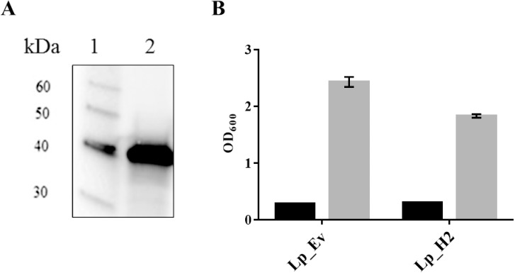
Fig 2.
Detection of anchor-fused H2 antigen (A) and cell growth of L. plantarum producing the H2 antigen (B). Panel A shows Western blot analysis of a cell-free protein extract from Lp_H2 (lane 2) and a molecular mass standard (1). The predicted molecular mass of the Lp_1261H2-DC protein is 37 kDa. Panel B shows the growth rate of Lp_H2 and Lp_Ev, used as a control. The OD600 was measured at the induction point (black bars) and 3 h after induction (gray bars) for both strains. The data are presented as means of triplicates ± SEM.

