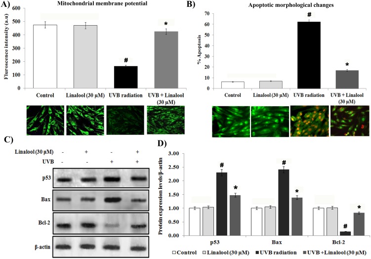
Fig 4. Linalool protects UVB-induced apoptosis in HDFa cells.
(A) Linalool prevents UVB induced alteration of mitochondrial membrane potential in HDFa cells. Linalool treated and/or UVB irradiated cells were treated with Rh-123 staining. Fluorescence microscopic images (20X) were recorded using fluoresece microscope (Cell imaging station, life technologies) under green fluorescence lamb. Bar diagram represents fluorescence intensity made with excitation and emission at 485 ± 10 and 530 ± 12.5 nm, respectively using a multimode reader (Teccan, Austria). Data expressed as means ± SD of six experiments in each group. Values not sharing a common marking (# and *) differ significantly at P < 0.05 (Duncan’s multiple-range test). (B) Linalool against UVB induced apoptotic morphological changes were measured by AO/EtBr staining. Linalool treated and/or UVB irradiated cells were treated with AO/EtBr staining, fluorescence microscopic images (20X) were recorded and % apoptotic cells were calculated. (C) Effect of linalool on UVB-induced expression of apoptotic markers in HDFa. Western blot analysis of Bax, p53 and Bcl-2 expression in HDFa. (D) Band intensities were analyzed by Image studio software and normalized to ß-actin level. Values are given as means ± SD of six experiments in each group. Values not sharing a common marking (# and *) differ significantly at P < 0.05 (DMRT).

