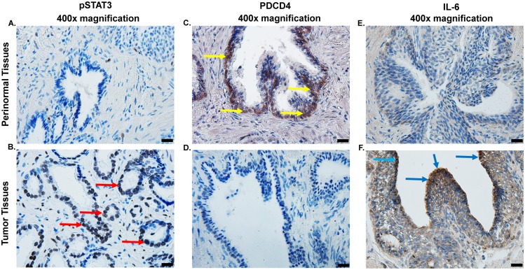
Fig 8. Levels of pSTAT3, PDCD4 and IL-6 in human prostate specimens.
(A, B) Immunohistochemistry studies were performed to determine the levels of pSTAT3 in formalin-fixed paraffin embedded prostate tumor specimens, as compared to adjacent normal prostate specimen. Diaminobenzidine tetrahydrochloride (DAB) staining revealed high expression of pSTAT3 (B) as dark-brown labeling in epithelial cells of tumor samples (marked by red arrows) while low expression was observed in paired perinormal prostate tissues (A). In contrast, high expression of PDCD4 was observed in perinormal prostate tissues (as evidenced by dark-brown labeling, indicated by yellow arrows) (C), as compared to prostate tumors (D). Immunolabling of prostate tumor revealed high expression of IL-6 (F), compared to perinormal prostate specimens. Representative images show immunohistochemical studies performed in four different specimens obtained from three different patients. Pictures magnification is 400x. Scale bar is 20 μm.

