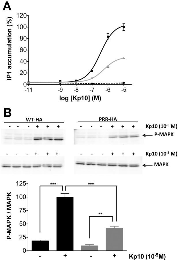
Figure 2.
The additional PRR disturbs the maximal Kp10-induced activation of KISS1R in HeLa cells. A, IP production in WT-HA-KISS1R (black line), PRR-HA-KISS1R (gray line), and pcDNA3.1-transfected cells (dotted line) induced by 1-hour incubation with Kp10. Data are shown as the mean ± SEM of the 2 experiments, each performed in triplicates. B, MAPK pathway activation by a 10-minute treatment with Kp10 at 10−5M. The density of Phospho-MAPK (P-MAPK) bands was quantified and normalized by the density of MAPK bands. Data are the mean of triplicates (WT-HA-KISS1R in black and PRR-HA-KISS1R in gray); **P < .01 and ***P < .001.

